CHAPTER 3: Senses and Perception
In this Chapter
Vision
1The wonderful sense of sight allows us to experience the world, from the genius of Michelangelo’s Sistine Chapel ceiling to the mist-filled vista of a mountain range. Vision is one of our most delicate and complicated senses. Many processes must occur simultaneously in order for us to see what is happening around us. Information about image size and shape, color, motion, and location in space all must be gathered, encoded, integrated, and processed. Performing these activities involves about 30 percent of the human brain — more than for any other sense.
2Vision has been studied intensively. As a result, neuroscientists may know more about it than any other sensory system. Most information about initial stages of visual transduction, or how light is converted into electrical signals, comes from studies of Drosophila (fruit flies) and mice, whereas visual processing has been mostly studied in monkeys and cats.
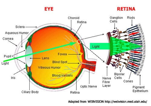
3It all Starts with Light. Vision begins with light passing to the cornea, which does about three-quarters of the focusing, and then the lens, which adjusts the focus. Both combine to produce a clear image of the visual world on a sheet of photoreceptors called the retina, which is part of the central nervous system but located at the back of the eye.
4Photoreceptors gather visual information by absorbing light and sending electrical signals to other retinal neurons for initial processing and integration. The signals are then sent via the optic nerve to other parts of brain, which ultimately processes the image and allows us to see.
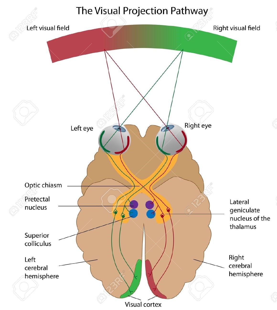
5As in a camera, the image on the retina is reversed: Objects to the right of center project images to the left part of the retina and vice versa; objects above the center project to the lower part and vice versa. The size of the pupil, which regulates how much light enters the eye, is controlled by the iris. The shape of the lens is altered by the muscles just behind the iris so that near or far objects can be brought into focus on the retina.
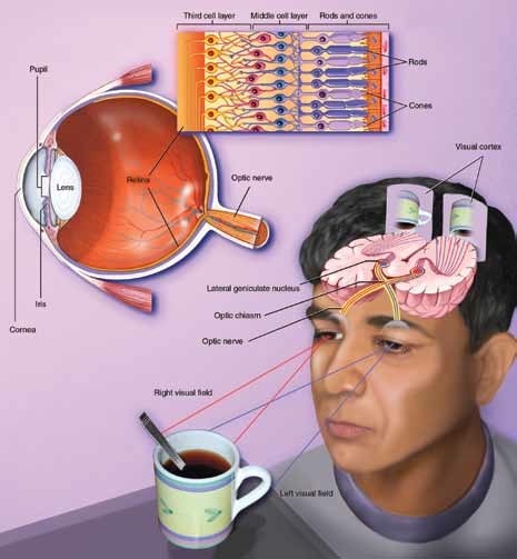
6Primates, including humans, have well-developed vision using two eyes, called binocular vision. Visual signals pass from each eye along the million or so fibers of the optic nerve to the optic chiasm, where some nerve fibers cross over. This crossover allows both sides of the brain to receive signals from both eyes.
7When you look at a scene with both eyes, the objects to your left register on the right side of the retina. This visual information then maps to the right side of the cortex. The result is that the left half of the scene you are watching registers in the cerebrum’s right hemisphere. Conversely, the right half of the scene registers in the cerebrum’s left hemisphere. A similar arrangement applies to movement and touch: Each half of the cerebrum is responsible for processing information received from the opposite half of the body.
8Scientists know much about the way cells encode visual information in the retina, but relatively less about the lateral geniculate nucleus — an intermediate way station between the retina and visual cortex — and the visual cortex. Studies about the inner workings of the retina give us the best knowledge we have to date about how the brain analyzes and processes sensory information.
9Photoreceptors, about 125 million in each human eye, are neurons specialized to turn light into electrical signals. Two major types of photoreceptors are rods and cones. Rods are extremely sensitive to light and allow us to see in dim light, but they do not convey color. Rods constitute 95 percent of all photoreceptors in humans. Most of our vision, however, comes from cones that work under most light conditions and are responsible for acute detail and color vision.
10The human eye contains three types of cones (red, green and blue), each sensitive to a different range of colors. Because their sensitivities overlap, cones work in combination to convey information about all visible colors. You might be surprised to know that we can see thousands of colors using only three types of cones, but computer monitors use a similar process to generate a spectrum of colors. The central part of the human retina, where light is focused, is called the fovea, which contains only red and green cones. The area around the fovea, called the macula, is critical for reading and driving. Death of photoreceptors in themacula, called macular degeneration, is a leading cause of blindness among the elderly population in developed countries, including the United States.
11The retina contains three organized layers of neurons. The rod and cone photoreceptors in the first layer send signals to the middle layer (interneurons), which then relays signals to the third layer, consisting of multiple different types of ganglion cells, specialized neurons near the inner surface of the retina. The axons of the ganglion cells form the optic nerve. Each neuron in the middle and third layer typically receives input from many cells in the previous layer, and the number of inputs varies widely across the retina. Near the center of the gaze, where visual acuity is highest, each ganglion cell receives inputs — via the middle layer — from one cone or, at most, a few, allowing us to resolve very fine details. Near the margins of the retina, each ganglion cell receives signals from many rods and cones, explaining why we cannot see fine details on either side. Whether large or small, the region of visual space providing input to a visual neuron is called its receptive field.
13How Visual Information Is Processed. About 60 years ago, scientists discovered that each vision cell’s receptive field is activated when light hits a tiny region in the center of the field and inhibited when light hits the area surrounding the center. If light covers the entire receptive field, the cell responds weakly. Thus, the visual process begins by comparing the amount of light striking any small region of the retina with the amount of surrounding light.
14Visual information from the retina is relayed through the lateral geniculate nucleus of the thalamus to the primary visual cortex — a thin sheet of tissue (less than one-tenth of an inch thick), a bit larger than a half-dollar, which is located in the occipital lobe in the back of the brain. The primary visual cortex is densely packed with cells in many layers, just as the retina is. In its middle layer, which receives messages from the lateral geniculate nucleus, scientists have found responses similar to those seen in the retina and in lateral geniculate cells. Cells above and below this layer respond differently. They prefer stimuli in the shape of bars or edges and those at a particular angle (orientation). Further studies have shown that different cells prefer edges at different angles or edges moving in a particular direction.
15Although the visual processing mechanisms are not yet completely understood, recent findings from anatomical and physiological studies in monkeys suggest that visual signals are fed into at least three separate processing systems. One system appears to process information mainly about shape; a second, mainly about color; and a third, movement, location, and spatial organization. Human psychological studies support the findings obtained through animal research. These studies show that the perception of movement, depth, perspective, the relative size of objects, the relative movement of objects, shading, and gradations in texture all depend primarily on contrasts in light intensity rather than on color. Perception requires various elements to be organized so that related ones are grouped together. This stems from the brain’s ability to group the parts of an image together and also to separate images from one another and from their individual backgrounds.
16How do all these systems combine to produce the vivid images of solid objects that we perceive? The brain extracts biologically relevant information at each stage and associates firing patterns of neuronal populations with past experience.
18Research Leads to More effective Treatment. Vision studies also have led to better treatment for visual disorders. Information from research in cats and monkeys has improved the therapy for strabismus, a condition in which the eyes are not properly aligned with each other and point in different directions. It is also termed squint, cross-eye, or walleye. Children with strabismus initially have good vision in each eye. But because they cannot fuse the images in the two eyes, they tend to favor one eye and often lose useful vision in the other. Vision can be restored in such cases, but only during infancy or early childhood. Beyond the age of 8 or so, the blindness in one eye becomes permanent. Until a few decades ago, ophthalmologists waited until children reached the age of 4 before operating to align the eyes, prescribing exercises, or using an eye patch. Now strabismus is corrected very early in life — before age 4 — when normal vision can still be restored.
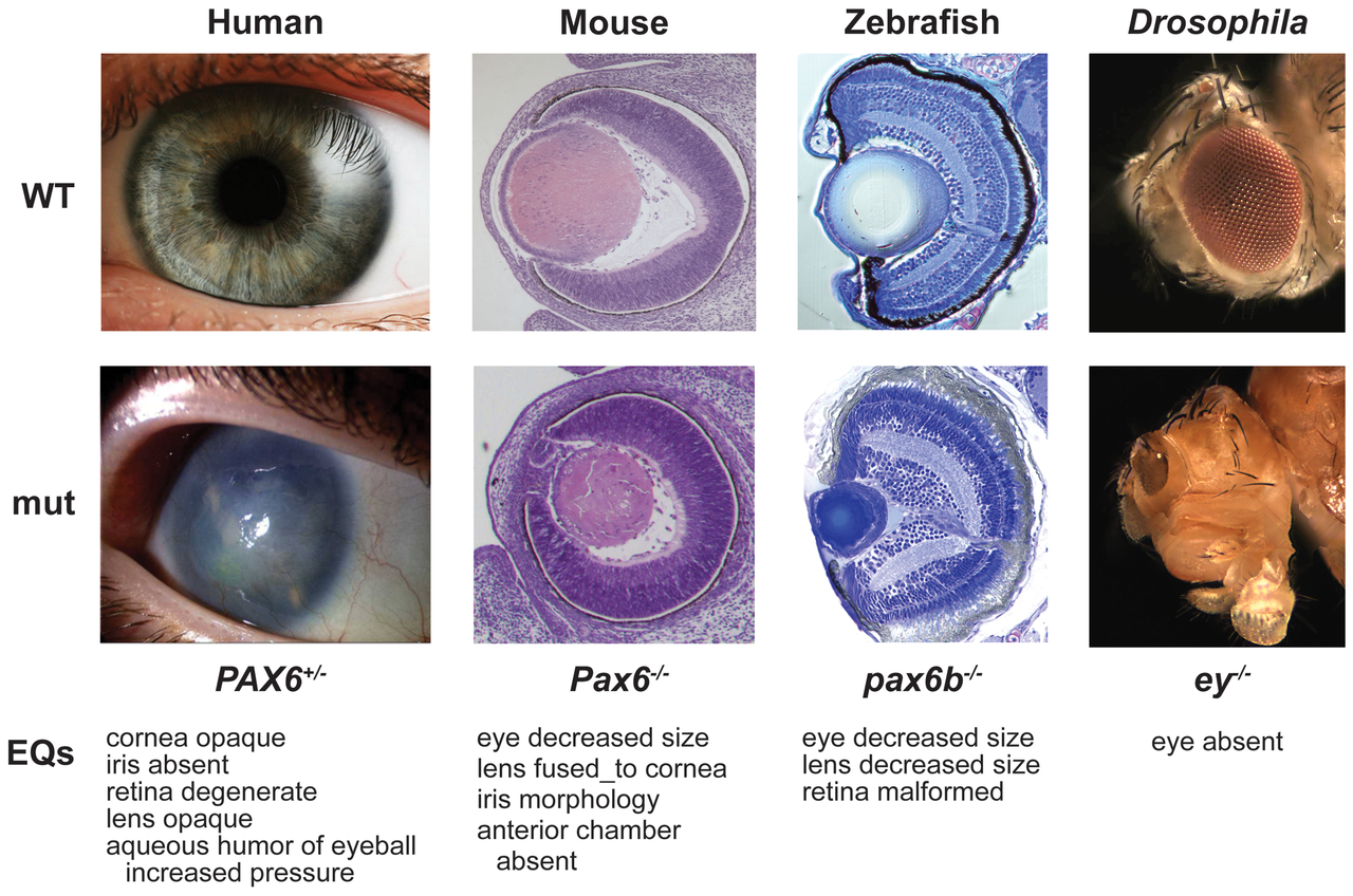
19Extensive genetic studies and use of model organisms have allowed us to identify defects in inherited eye diseases, making it possible to design gene or stem cell-based therapy and discover new drugs for treatment. Loss of function or death of photoreceptors appears to be a major cause of blindness in many diseases that are currently incurable. Recently, gene therapy for a small group of patients with severe blindness allowed them to see. Work also is in progress to bypass lost photoreceptors and send electrical signals directly to the brain via ganglion cells.