CHAPTER 1: Brain Basics
Anatomy of the Brain and the Nervous System
1The brain is the body’s control center, managing just about everything we do. Whether we’re thinking, dreaming, playing sports, or even sleeping, the brain is involved in some way. The brain is organized into different parts that are wired together in a specific way. Each part has a specific job (or jobs) to do, making the brain the ultimate multitasker. Working in tandem with the rest of the nervous system, the brain sends and receives messages, allowing for ongoing communication.
2Mapping the Brain The cerebrum, the largest part of the human brain, is associated with higher order functioning, including the control of voluntary behavior. Thinking, perceiving, planning, and understanding language all lie within the cerebrum’s control. The cerebrum is divided into two hemispheres — the right hemispheres and the left hemisphere. Bridging the two hemispheres is a bundle of fibers called the corpus callosum. The two hemispheres communicate with one another across the corpus callosum.
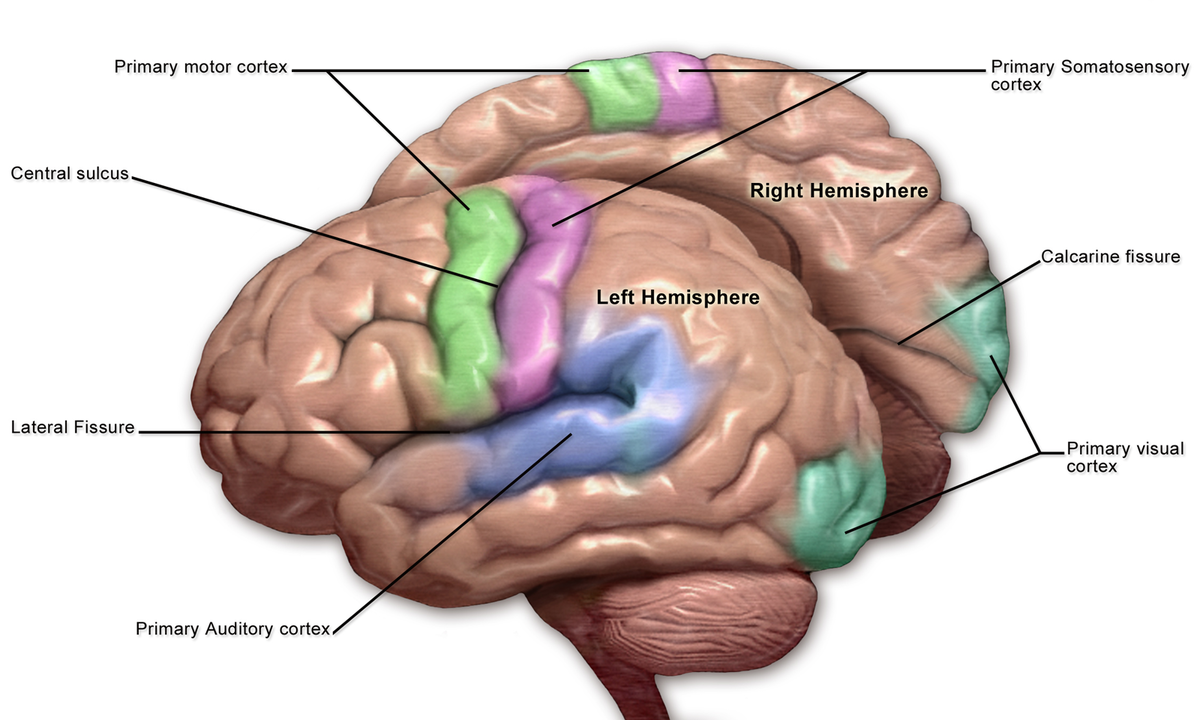
3Covering the outermost layer of the cerebrum is a sheet of tissue called the cerebral cortex. Because of its gray color, the cerebral cortex is often referred to as gray matter. The wrinkled appearance of the human brain also can be attributed to characteristics of the cerebral cortex. More than two-thirds of this layer is folded into grooves. The grooves increase the brain’s surface area, allowing for inclusion of many more neurons.
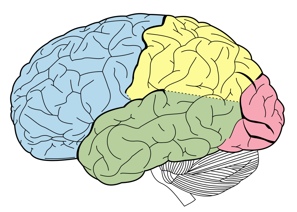
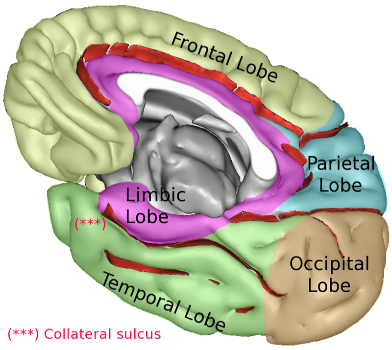
4The function of the cerebral cortex can be understood by dividing it somewhat arbitrarily into zones, much like the geographical arrangement of continents.
5The frontal lobe is responsible for initiating and coordinating motor movements; higher cognitive skills, such as problem solving, thinking, planning, and organizing; and for many aspects of personality and emotional makeup.
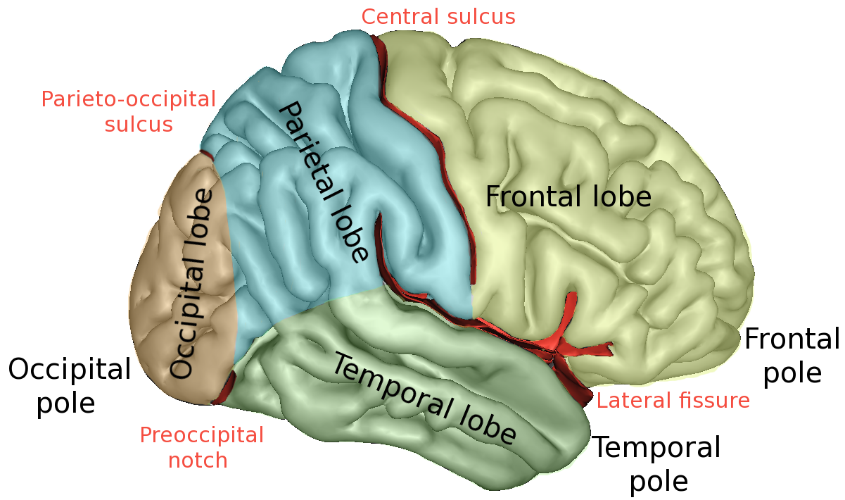
6The parietal lobe is involved with sensory processes, attention, and language. Damage to the right side of the parietal lobe can result in difficulty navigating spaces, even familiar ones. If the left side is injured, the ability to understand spoken and/or written language may be impaired.
7The occipital lobe helps process visual information, including recognition of shapes and colors.
8The temporal lobe helps process auditory information and integrate information from the other senses. Neuroscientists also believe that the temporal lobe has a role to play in short-term memory through its hippocampal formation, and in learned emotional responses through its amygdala.
9 All of these structures make up the forebrain.
10Other key parts of the forebrain include the basal ganglia, which are cerebral nuclei deep in the cerebral cortex; the thalamus ; and the hypothalamus. The cerebral nuclei help coordinate muscle movements and reward useful behaviors; the thalamus passes most sensory information on to the cerebral cortex after helping to prioritize it; and the hypothalamus is the control center for appetites, defensive and reproductive behaviors, and sleep-wakefulness.
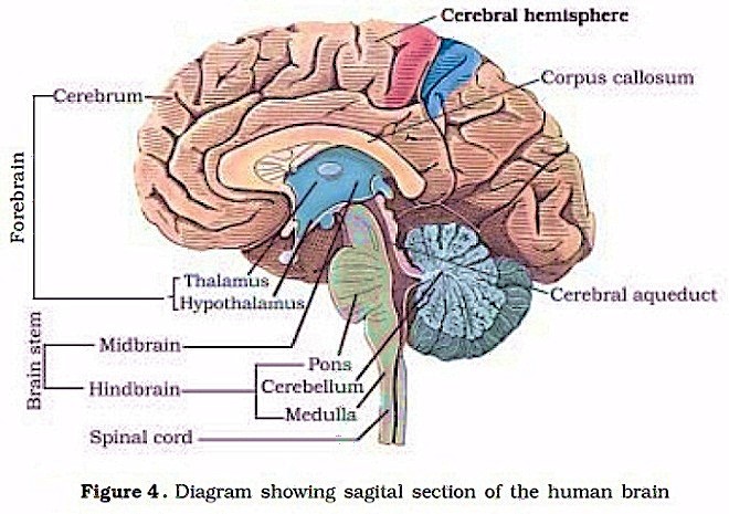
11The midbrain consists of two pairs of small hills called colliculi. These collections of neurons play a critical role in visual and auditory reflexes and in relaying this type of information to the thalamus. The midbrain also has clusters of neurons that regulate activity in widespread parts of the central nervous system and are thought to be important for reward mechanisms and mood.
12The hindbrain includes the pons and the medulla oblongata, which control respiration, heart rhythms, and blood glucose levels.
13Another part of the hindbrain is the cerebellum which, like the cerebrum, also has two hemispheres. The cerebrum’s two hemispheres help control movement and cognitive processes that require precise timing, and also play an important role in Pavlovian learning.
14The spinal cord is the extension of the brain through the vertebral column. It receives sensory information from all parts of the body below the head. It uses this information for reflex responses to pain, for example, and it also relays the sensory information to the brain and its cerebral cortex. In addition, the spinal cord generates nerve impulses in nerves that control the muscles and the viscera, both through reflex activities and through voluntary commands from the cerebrum.
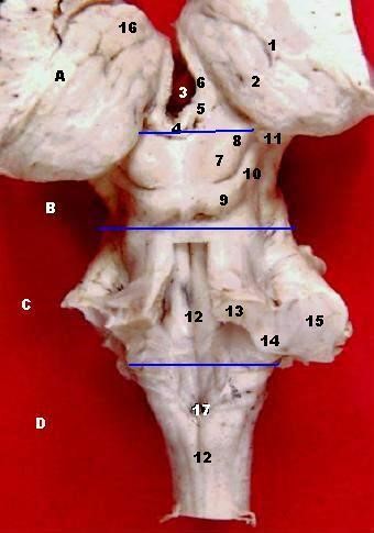
(6) Human brainstem and thalamus - posterior view
- Brachium colliculi inferioris
- Corpus geniculatum mediale
- Sulcus medianus
- Pedunculus cerebellaris superior
- Pedunculus cerebellaris inferior
- Pedunculus cerebellaris medius
- Tuberculum anterius thalami
- Obex, Area postrema
- Taenia choroidea (and lateral: Lamina affixa, Stria terminalis)
- Thalamus, Pulvinar thalami
- (Ventriculus tertius)
- Stalk of Glandula pinealis
- Habenula
- Stria medullaris
- Colliculus superior
- Brachium colliculi superioris
- Colliculus inferior
A: Thalamus B: Mesencephalon C: Pons D: Medulla oblongata
On this specimen, the following thalamic structures can be seen: 1. the Epithalamus (Stria Medullaris Thalami, Habenula, & Pineal), 2. the Anterior Nucleus of the dorsal thalamus (Anterior Tubercle) and, 3. the Pulvinar (the large posterior portion of the dorsal thalamus which overhangs the midbrain. The Medulla, Pons & Midbrain are delineated on the posterior surface of the brainstem.
NOTE: The 4 Colliculi of the tectum are referred to collectively as the Quadrigeminal Plate. The three Cerebellar Peduncles are shown here as they enter the brainstem on each side. In the Midbrain identify the Superior Colliculus and Inferior Colliculus. Also identify the Brachium of the Superior Colliculus and the Brachium of the Inferior Colliculus which connect with the Lateral Geniculate Body and Medial Geniculate Body, respectively.
The cerebellum forms the roof of the 4th ventricle and is connected to the brainstem by 3 pairs of peduncles or pillars (shown on right side of brainstem) . The peduncles are made up of axons entering and leaving the cerebellum. The Inferior Cerebellar peduncle projects from the medulla, the large Middle Cerebellar Peduncle projects from the Pons, and the Superior Cerebellar Peduncle connects with the midbrain.
15The Parts of the Nervous System The forebrain, midbrain, hindbrain, and spinal cord form the central nervous system (CNS), which is one of two great divisions of the nervous system as a whole. The brain is protected by the skull, while the spinal cord, which is about 17 inches (43 cm) long, is protected by the vertebral column.
16The other great division of the human brain is the peripheral nervous system (PSN), which consists of nerves and small concentrations of gray matter called ganglia, a term specifically used to describe structures in the PNS. Overall the nervous system is a vast biological computing device formed by a network of gray matter regions interconnected by white matter tracts.
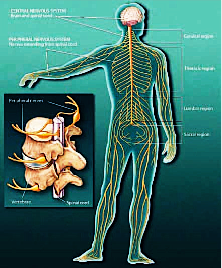
17The brain sends messages via the spinal cord to peripheral nerves throughout the body that serve to control the muscles and internal organs. The somatic nervous system (SNS) is made up of neurons connecting the CNS with the parts of the body that interact with the outside world. Somatic nerves in the cervical region are related to the neck and arms; those in the thoracic region serve the chest; and those in the lumbar and sacral regions interact with the legs.
18The autonomic nervous system (ANS) is made of neurons connecting the CNS with internal organs. It is divided into two parts. The sympathetic nervous system mobilizes energy and resources during times of stress and arousal, while the parasympathetic nervous system conserves energy and resources during relaxed states, including sleep.
19Messages are carried throughout the nervous system by the individual units of its circuitry: neurons. The next section describes the structure of neurons, how they send and receive messages, and recent discoveries about these unique cells.